FRIDAY, 11 MAY 2012
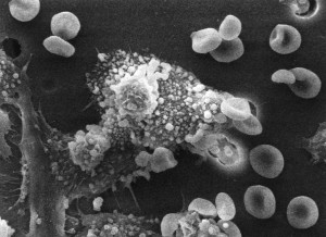
The nanoparticles themselves are actually gold stars, approximately 25 nanometres in width. They have been described by Teri Odom, who led the study on human cervical and ovarian cancer cells as “tiny hitchhikers”. This is because the stars are attracted to a protein on the surface of the cancer cell which then “conveniently shuttles the nanostars to the cell’s nucleus”. Upon reaching the nucleus the drug is released from the surface of the nanostar and starts targeting the cancer.The research carried out by Odom and her team is also impressive as they have been the first to image how the nanoparticles interact with a cancer cell’s nucleus. Using electron microscopy the team observed how the drug-loaded nanoparticles radically changed the shape of the cancer cell nucleus from a smooth ellipsoid to a deformed, uneven shape with deep folds. This change was due to cells dying and the cell population becoming less viable –both very good news for cancer treatment.
Since the initial research the nano-hitchikers have had similar effects on twelve other types of human cancer cell lines, suggesting the development could lead to generalised treatment for different cancers. The large surface are of the nanostars are a very efficient method of drug delivery, as a high concentration of drug molecules can be loaded onto the star.
Nanostar development seems to have advanced several important areas in cancer treatment and drug delivery design, with positive and valuable results. In years to come people may well be thanking their nanostars.
Written by Laura Stevens
DOI: 10.1021/nn300296p

