WEDNESDAY, 15 MAY 2013
In the 1670s Antonie Van Leeuwenhoek revolutionised science when he began to experiment with magnification. His curiosity to observe anything that could be placed under a magnifying lens led him to be the first person to describe many microscopic entities, such as bacteria, which he isolated from his own tooth plaque, algae, nematode worms, and sperm. Leeuwenhoek’s discoveries rightly earned him the title of the ‘Father of Microbiology’, and his descriptions of the fascinating microscopic detail of the world fuelled the development of microscopes like those still in use today.Since the time of Leeuwenhoek and his contemporaries, we have been trying to engineer microscopes that will allow us to view increasingly small objects in ever more detail. Optical or light microscopes, like those you might see in any lab or science classroom, use light and a system of lenses to magnify small samples, allowing us to view them as if they were up to 2000 times larger. However, the wavelength of visible light limits these light microscopes in their resolution to around 200 nanometres (200 billionths of a meter); two objects that are closer together than this cannot be distinguished any more. While this is sufficient to clearly see plant and animal cells as well as bacteria, it is not suitable to study organelles within the cell, such as the nucleus, mitochondria or chloroplasts, or to see most viruses.
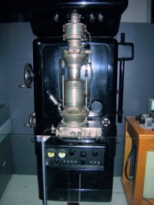
In the 1920s it was shown that accelerated electrons could behave like light waves when in a vacuum, and furthermore, the path of these electrons could be shaped by electric and magnetic fields in a similar way to how glass lenses focus light. In 1933, these discoveries lead the German scientist Ernst Ruska to build the first microscope that used electrons rather than light waves. It was named, rather unimaginatively, the ‘electron microscope’.
Ruska won the Nobel Prize for his work, but not until 1986. Initially, his invention didn’t impress the scientific community as it was impractical to use, had the tendency to burn samples and its resolution was no better than that of traditional light microscopes. It wasn’t until five years and several prototypes after his first one that the electron microscope gained popularity. This was in no small part due to the discovery that coating biological samples with heavy metals, such as lead or uranium, helped separate electrons, thus giving better contrast.
Electron microscopes magnify by firing streams of electrons at an object. The electrons hit the object, bounce off and are focused by electromagnetism onto a screen or a photographic plate to make a visible image. As an electron has a wavelength around 100,000 times shorter than a visible light wave, the electron microscope has a much greater resolving power and can be used to reveal the structure of much smaller objects than can be seen with light microscopes. Modern electron microscopes can resolve something as small as 50 picometers (around one trillionth of a meter) and magnify it by up to 10 million times. This resolution is staggeringly high when you consider that the diameter of a hydrogen atom is just 100 picometers. Such high resolution allows scientists to study cellular compartments and gain a greater understanding of what goes on inside a cell.
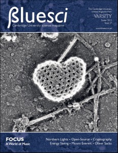
This issue’s cover image illustrates the incredible detail that can be achieved when using electron microscopy. Taken by Daniela Sahlender from Margaret Robinson’s lab in Clinical Biochemistry, the image shows a sub-cellular structure called a clathrin-coated vesicle, which has been magnified 1.6 million times. Clathrin vesicles are small bubbles, around 100-200 nanometers in size that are used to transport molecules, such as nutrients and hormones within and between cells. The vesicles are able to pass from the outside of a cell into the cell, a process known as endocytosis. The vesicles have a membrane similar to the outer membrane of a cell and these two membranes can merge, allowing the content of the vesicle access to the inside of the cell. The cover image shows a clathrin-coated vesicle undergoing endocytosis and budding out from the cell membrane.
Antonie Van Leeuwenhoek would never have believed that his work with lenses would lead to the discovery of microscopes so powerful that tiny intracellular structures could clearly be seen. As we continue to build ever more powerful microscopes, who knows what else we might discover about the microscopic world around us.
Nicola Love is a 3rd year PhD student at the Department of Physiology, Development and Neuroscience


