MONDAY, 3 FEBRUARY 2014
Every single organism, no matter how large or complex, began its existence as a single cell. It is truly startling to think that we were all once about the same size as the full stop at the end of this sentence. From this tiny, microscopic fertilised egg (known as a zygote in mammals) we are able to divide and develop and form every single organ and tissue in the body. Understanding this process, how a single cell is able to pattern itself into a complex organism, has been the subject of a staggering amount of research over a number of decades. This is at the very heart of developmental biology. We want to know how one cell develops into millions of different organised structures.The very first pattering events in mammalian embryos occur over the first few days of development. During this time it is critical that the embryo establishes the correct morphological and molecular events necessary to allow the embryo to implant into the uterine wall. Once the embryo is adhered to the uterus, the mother is able to provide oxygen and nutrients to the developing foetus while at the same time removing waste products and carbon dioxide. The pre-implantation period of the embryo is thus a short but critical stage of development. In order for implantation to occur, the cells of the embryo need to spatially segregate and differentiate into three distinct cell lineages with the differing abilities to become specific tissues. This spatially segregated structure is known as a blastocyst and occurs through two cell fate decisions. The first cell fate decision gives rise to the trophectoderm, a supporting tissue which will go on to form the embryonic portion of the placenta, and the inner cell mass. The placenta connects the developing foetus to the uterine wall. The second cell fate decision is responsible for segregating the inner cell mass into the pluripotent epiblast, which will go on to form all the cells of the embryo proper, and the second supporting tissue, the primitive endoderm, which gives rise to yolk sac which acts as a circulatory system for the foetus.
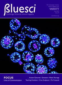
The image on the cover of this issue of BlueSci shows a group of cultured mouse embryos that have developed until the blastocyst stage, just before implantation into the uterine wall. It was taken by Mubeen Goolam in Professor Zernicka-Goetz’s lab in The Gurdon Institute at the University of Cambridge. The embryos were stained with fluorescent markers capable of binding different cell-type specific proteins which, in turn, allows the identification of different cell lineages present in the blastocyst. The blue cells are the trophectoderm, the white cells are the epiblast, and the pink cells are the primitive endoderm. The image also shows that a number of the blastocysts appear to have ‘burst’ and have regions where the cells are ‘spilling out’ of their original boundary. This is expected and is not an artefact of the staining procedure. These blastocysts are ready to implant into the uterine wall and thus have burst out of their membranous boundary. This is evidence that they have developed perfectly while in culture.
Mice are the perfect model to use to study mammalian development as their small size, short gestation period, and the ease of handling them allows one to do a large volume of research in relatively efficient amount of time and space. Additionally, mice are highly similar to humans on a genetic and physiological level and have a very well documented genome that is easy to manipulate and analyse. We can thus use mice to understand human development.
While mice as a model organism have existed for some time, it is the recent developments in microscopy and our ability to culture these embryos in the laboratory that have allowed us to probe deeper into the events that occur during early mammalian development. We now have the ability to create high resolution time lapse images that allow one to visualise the very process of development as it occurs. We can literally watch development as it takes place, something which would have been impossible to imagine a few years ago.
Our ability to mark and identify cells with different cell fates means that we can create images that are not only visually beautiful, but that have biological meaning too. The advancements in technology we have seen in a short space of time will allow us to explore and examine the very first stages of development in a way that would have been impossible only a few years ago. Who knows what we might discover?
Mubeen Goolam’s image was one of three winning entries in the University of Cambridge Graduate School of Life Sciences (GSLS) image competition. The competition, run during the Cambridge Science Festival, showcases the variety of biological research in Cambridge. To find out more, visit the GSLS website. The other winning images, taken by Nuri Purswani and Xana Almeida, can be found on www.bluesci.org.


